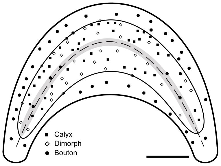
Figure 3.
Schematic illustration of lagenar apical surface with the locations of all 114 reconstructed afferents. The striola (thin black outline) was defined by the area containing calyx terminals. The extrastriola exclusively contained bouton afferents (circles). The reversal line (italics) ran through the central epithelium and was flanked by the Type II band (gray shade). Scale bar = 200μm.

