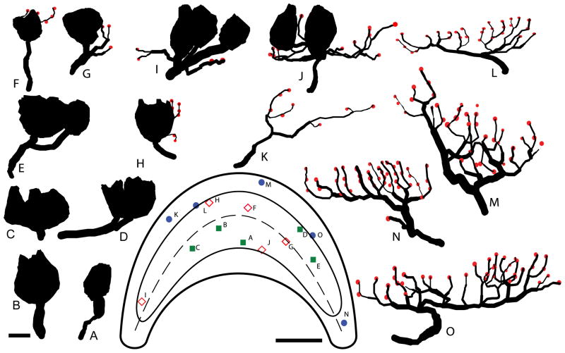
Figure 4.
Innervation patterns of calyx, dimorph, and bouton afferents. Reconstructions of 5 calyx (A–E), 5 dimorph (F – J), and 5 bouton (K – 0) afferents are arranged in order of increasing complexity. Calyx terminals (closed contours) and bouton terminals (red circles) are shown, with all reconstructions drawn to scale. Each afferent’s corresponding location in the lagena (inset) is indicated on the illustrated apical surface. Scale bar = 5μm for the reconstructions; 200μm for the surface.

