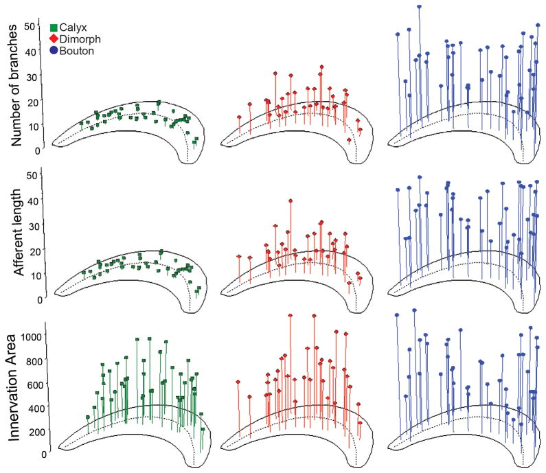
Figure 6.
Number of branches, afferent length and innervation area for fiber types. The locations of calyx (green squares; left panels), dimorph (red diamonds; middle panels) and bouton (blue circles; right panels) afferents are shown as a function of number of branches (tip), afferent fiber length (middle, μm), and innervation area (bottom, μm2), respectively. Dashed line represents reversal line.

