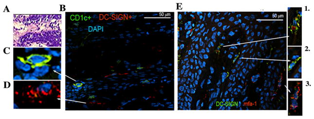
FIGURE 4.
P. gingivalis infected mDCs in oral submucosa of CP patient A. Representative gingival tissue section (7μM thick) from a CP patient stained with hematoxylin and eosin (H&E) (20x). B. Section was stained with FITC-conjugated mouse anti-human CD1c (BDCA-1) and RPE-conjugated mouse anti-human CD209 (DC-SIGN) and image captured at 100x. C. Shown at ~500x final enlargement (optical and digital) is detail view of individual of CD1c+ mDC/LC in epithelium and D. DC-SIGN+ mDCs in lamina propria (200x). E. FITC-conjugated mouse anti-human CD209 and Alexa Fluor 594 conjugated to mfa-1 antibody AEZαMfa1 using commercial DyLight™ microscale antibody labeling kit in CP oral mucosal tissues (100x). Shown in panels 1–3 (at 200x) are detail of areas of mfa-1-DC-SIGN colocalization.

