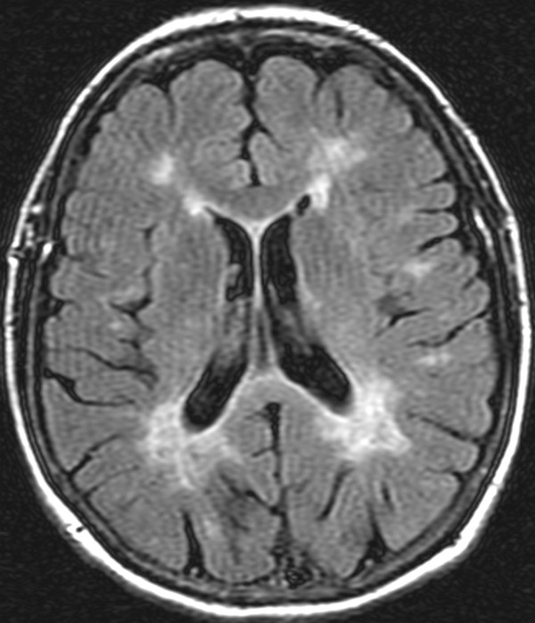
Figure 1.
Magnetic resonance imaging scans: fluid attenuated inversion recovery (FLAIR) sequence, transverse plane. Shown are demyelinating areas involving the periventricular areas of the lateral ventricles, more prominent in the posterior. A similar demyelination process is present in the white matter of the brain sulci (medullary white matter).

