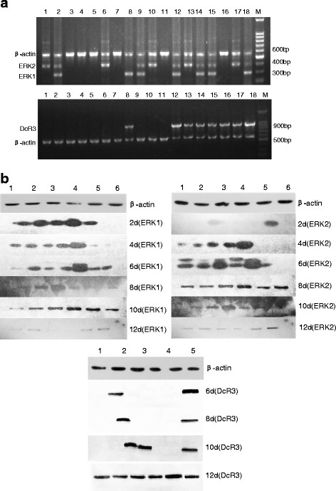
Figure 4.
The expression of DcR3 and ERK1/2 in mouse models analyzed by RT-PCR (Figure 4a) and Western blotting (Figure 4b). a: Lane M, DNA marker; lanes1-6, hearts, livers, spleens, lungs, kidneys and tumor tissues of mouse models on the fourth day; lanes7-12, which on the eighth day ; lanes13-18, on the twelfth day; ERK1 mRNA was positive in lane2,8,9,12,14,15,18, ERK2 mRNA was positive in lane1,2,6,9,10,12,13,14,15,17,18. DcR3 mRNA was detected in lane8, 12, 13, 14, 15, 16, 17, 18. b: lane 1, heart; lane2, liver; lane3, spleen; lane4,lung; lane5, kidney; lane6, tumor tissues; The expression of ERK1 protein increased in tumor tissues as time went on, in heart, liver and kidney persisted for 12 days, The expression of ERK2 could be detected in spleen and tumor from the second day which was positive in tumor tissues till the fourth day and all of five organs to the twelfth day. The expression of DcR3 protein was positive in tumor tissues and liver on the sixth day and in spleen on the tenth day, which was negative in heart, lung, kidney until the twelfth day.

