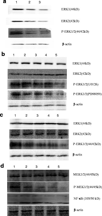
Figure 5.
The expression of ERK1/2 and P-ERK1/2 in BGC823 cell line with plasmid interference and inhibitor detected by Western blotting. a: The expression levels of ERK1/2 and P-ERK1/2 declined compared with the control. 1) control group; 2) 5:2 (plasmid: reagent); 3) 6:2 (plasmid: reagent). b: ERK1/2 phosphorylation gradually declined as the concentrations of the inhibitor U0126, PD98059 increased, but total ERK1/2 protein expression hardly changed. 1) 0 μmol/L; 2) 5 μmol/L; 3) 10 μmol/L; 4) 20 μmol/L; 5) 40 μmol/L. c: ERK1/2 phosphorylation gradually declined as the concentrations of the inhibitor APDC. 1) 0 μmol/L; 2) 10 μmol/L; 3) 20 μmol/L; 4) 40 μmol/L; 5) 80 μmol/L. d: PD98059 can obviously inhibit the MEK1/2 phosphorylation level, but it did not alter MEK1/2 or NF-κB expression levels. 1) 0 μmol/L; 2) 5 μmol/L; 3) 10 μmol/L; 4) 20 μmol/L; 5) 40 μmol/L.

