Figure 3.
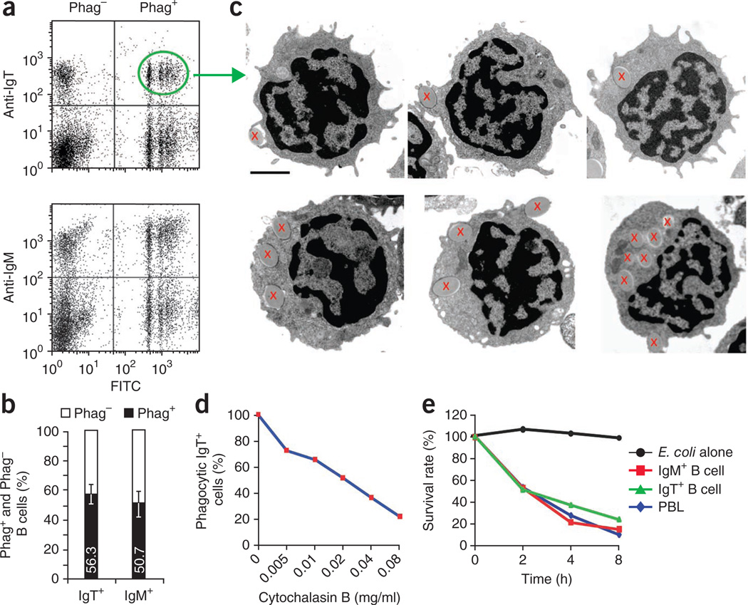
Phagocytic and intracellular killing capacities of IgT+ B cells. (a) Flow cytometry of peripheral blood leukocytes incubated with 1-µm fluorescent latex beads (labeled with fluorescein isothiocyanate (FITC)) and then stained with mAb to trout IgT or IgM (n = 9 fish). Phag−, nonphagocytic; Phag+, phagocytic. (b) Phagocytic and nonphagocytic cells in IgT+ or IgM+ B cell subsets of peripheral blood leukocytes (n = 9 fish). Numbers in bars indicate mean percent phagocytic cells. (c) Transmission electron microscopy of various stages of ingestion of 1-µm beads (red ‘x’) by phagocytic IgT+ B cells from peripheral blood leukocytes. Scale bar, 2 µm. (d) Inhibitory effect of cytochalasin B on the phagocytic capacity of IgT+ B cells, presented as the percentage of phagocytic cells relative to that of PBS-treated control cells. (e) Intracellular bacterial killing by sorted IgM+ and IgT+ B cells and total peripheral blood leukocytes incubated with live E. coli and lysed; lysates were inoculated onto Luria-Bertani agar plates and surviving intracellular bacteria were counted. Results are presented as percent of live bacteria at time 0, set as 100%. Data are representative of at least three independent experiments (mean and s.e.m. in b).

