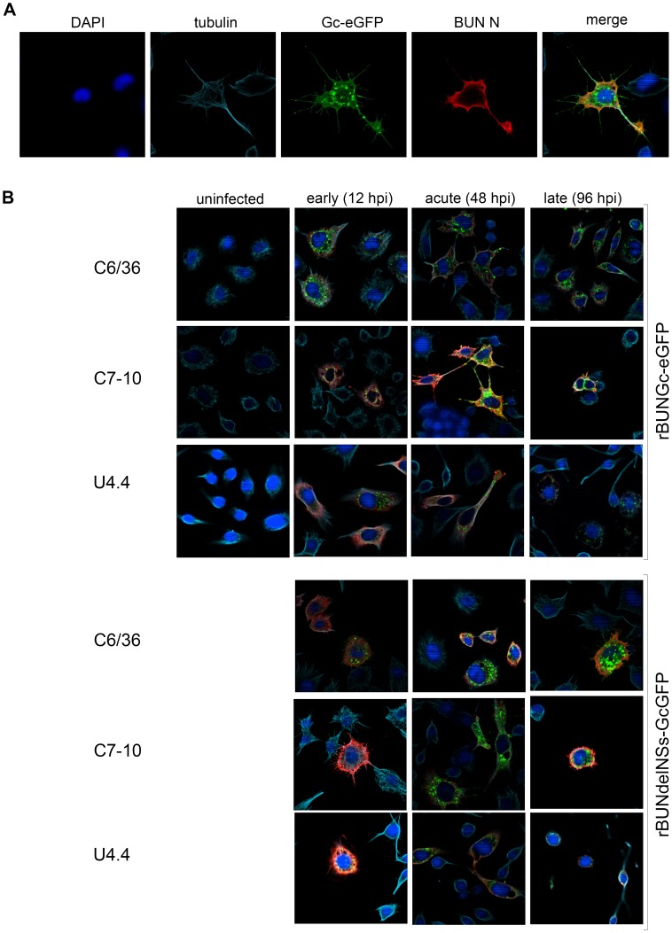
Figure 3. Immunofluorescence analysis of morphological changes in Ae. albopictus cell lines.
Mosquito cells were infected with rBUN-GcGFP or rBUNdelNSs-GcGFP at an MOI of 3 PFU/cell. The results present cells stained with anti-BUNN/anti-rabbit TexasRed (red signal), and with anti-tubulin/anti-mouse CY5 (light blue signal). The green signal shows autofluorescence of GFP-tagged Gc and the blue signal is DAPI staining of nuclei. A. C6/36 cell infected with rBUN-GcGFP virus during the acute stage of infection. B. C6/36, C7-10 and U4.4 cells were infected with fluorescent viruses. Three morphological stages of infection are presented: early, acute, and late. Only the merged images are shown.

