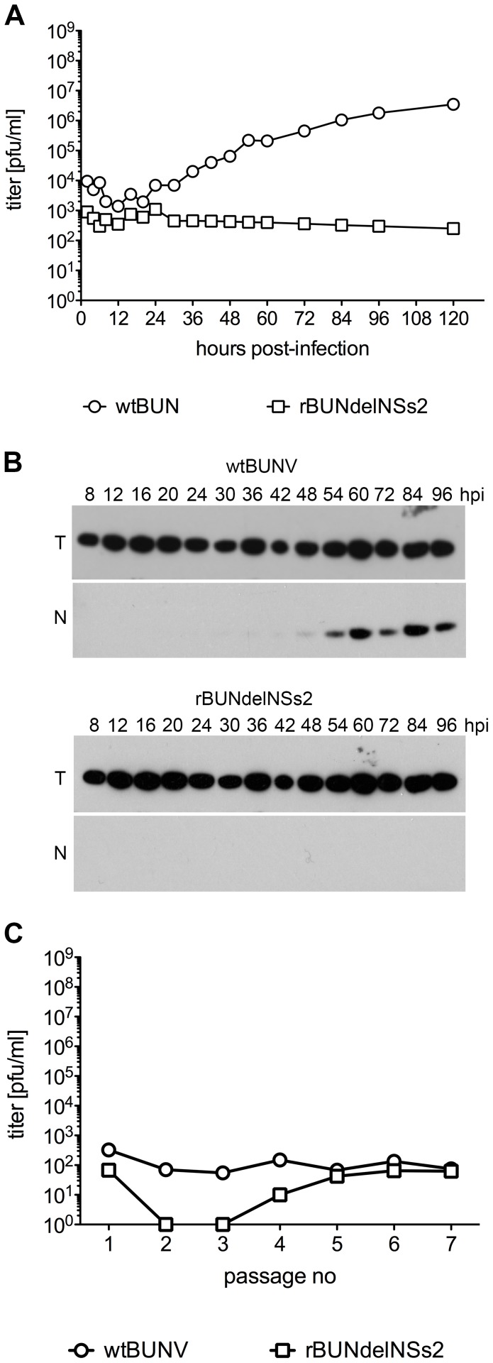
Figure 6. BUNV replication in Ae aegypti Ae cells.
A. Growth curves. Ae cells were infected with wtBUNV or rBUNdelNSs2 at an MOI of 1 PFU/cell. At the indicated times post infection supernatants were collected and assayed for the presence of infectious virus by plaque titration. B. Viral N protein expression. Cell extracts were prepared at the indicated times (h P.I.), separated by 12% SDS-PAGE, and proteins transferred to a membrane. The membrane was incubated with anti-BUNV N protein and tubulin (T) antibodies. C. Establishment of persistent infection. The titres of infectious virus in supernatants collected at each passage of infected cells were determined by plaque assay.

