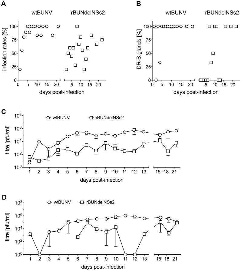
Figure 8. Virus replication in mosquito midguts and salivary glands.
Following infection via blood-meal, 9 engorged mosquitoes were collected at days 1 to 13, and 6 engorged mosquitoes were collected at days 15, 18 and 21. Midguts and salivary glands were dissected, pooled into groups of 3 organs, and infectious virus determined by plaque assay. A. Midgut infection rates. The percentage of virus positive midguts over total number of tested mosquitoes was calculated. B. Average virus titre per mosquito midgut. Error bars show the standard error between the different pools. C. Disseminated infection rates. The percentage of virus positive salivary glands over total number of positive mosquito midguts was calculated. D. Average virus titres per mosquito salivary gland. Error bars show the standard error between the different pools.

