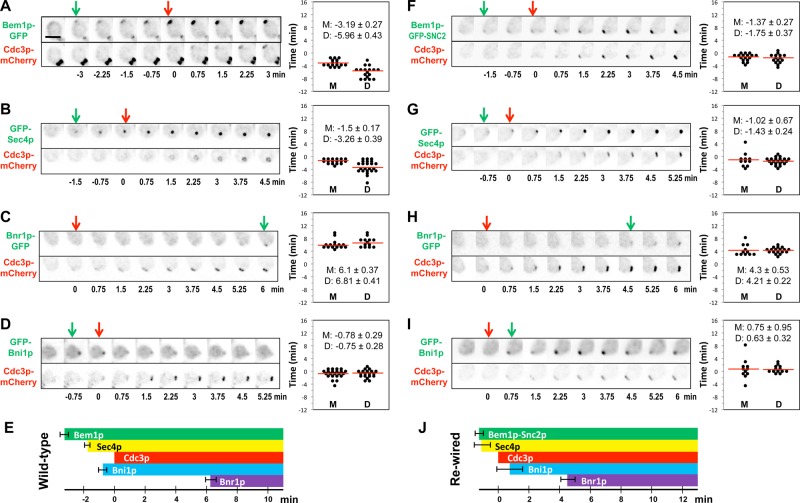
FIGURE 1:
Timing of protein polarization in wild-type and rewired cells. Wild-type (A–D) (DLY11909, 13890, 13344, 14014) and rewired (F–I) (DLY13072, 13884, 13945, 14026) cells were imaged and scored for the first appearance (arrows) of the indicated proteins at the incipient bud site. Inverted images are shown. Time intervals between polarization of GFP-tagged proteins and the septin Cdc3p-mCherry were quantified in mother and daughter cells (right; D, daughters; M, mothers). (E, J) Summary of the mean ± SEM timing in mother cells. Scale bar, 5 μm.

