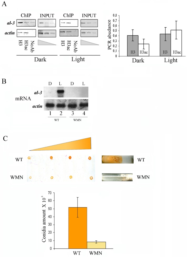
FIGURE 3:
Impairment of light-mediated H3 acetylation in the WMN strain. (A) ChIP assay on Neurospora WMN mycelia grown for 3 d in the dark before harvesting (Dark) or exposed to saturating white light for 5 min and then returned to the dark for 20 min before harvesting (Light 25 min). DNA immunoprecipitated with the anti–histone H3 (H3) and anti–acetyl histone H3 (H3ac) antibodies was amplified for the al-3 light-responsive promoter region. A sequence for actin was used as negative control. INPUT, PCR on chromatin samples before immunoprecipitation. ChIP, PCR from anti–hGCN5 IP samples. Noab, PCR from samples without antibody. Quantification of the PCR abundance for the al-3 promoter was calculated as described in Materials and Methods (right). The data are the mean ± SEM from three independent experiments. (B) Northern blot analysis of al−3mRNA in Neurospora WT and WMN strains kept in the dark (Dark) or after photoinduction (Light 25 min). The membrane was rehybridized using a probe against the actin mRNA as loading control (bottom). (C) WT and WMN strains were grown in slant for 4 d in the dark, illuminated for 5 h, and then kept in the dark for 8 h at 4°C to accumulate carotenoids. Increasing amounts of the conidia resuspended in water (5, 10, 20, and 25 μl) were spotted on a nitrocellulose filter to observe the increment of the red color corresponding to carotenoid content (top). Conidia count in WT and WMN was measured using a Burker cell counter (bottom). The data are the mean ± SEM from three independent experiments.

