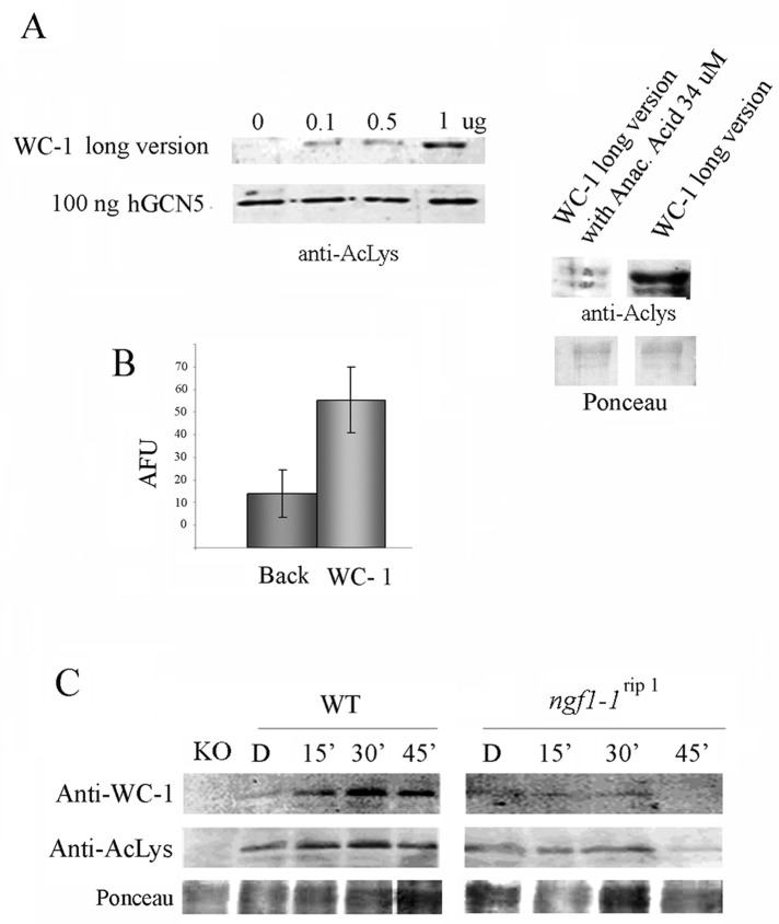
FIGURE 5:
WC-1 is an acetylated protein. (A) Progressive amounts of GST–WC-1 long version (Figure 2A) were tested in an in vitro acetylation assay in the presence of human GCN5, as described in Materials and Methods, using an anti–Pan acetylated-lysine antibody. Anacardic acid, a common HAT inhibitor, was used to prove the dependence of the acetylated signal on the acetyltransferase reaction. Ponceau staining was used to check the presence of similar WC-1 amounts on the membrane (right). (B) WC-1 acetylation was confirmed by a fluorescence in vitro acetylation assay. The data are the mean ± SEM from three independent experiments. AFU, arbitrary fluorescence units. (C) Western blot analysis with anti–WC-1 and anti–Pan acetylated- lysine antibodies of WT and ngf-1 RIP strains grown for 3 d in the dark before harvesting (Dark) or exposed to saturating white light for 5 min and then returned to the dark for 10, 25, and 55 min before harvesting (Light 15, 30, 60 min). A wc-1–null strain (KO) was used as a control of the specificity of the anti–WC-1 antibody. Ponceau staining was used to check the presence of similar WC-1 amounts on the membrane.

