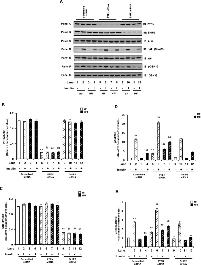
FIGURE 1:
Effect of PTEN or SHIP2 down-regulation on impaired Akt and GSK3β phosphorylation. Scrambled or PTEN- or SHIP2-specific siRNA–transfected N2A cells were differentiated in the absence (MF) or chronic presence (MFI) of insulin for 3 d and stimulated with or without insulin (100 nM) for 30 min. (A) Cell lysates were subjected to Western immunoblotting and probed with anti-PTEN (panel A), anti-SHIP2 (panel B), anti-actin (panel C), anti–phospho-Akt (Ser-473; panel D), anti-Akt (panel E), anti-phospho GSK3β (Ser-9; panel F), or anti-GSK3β (panel G) antibodies. (B) Bar diagram representing relative densitometric values of PTEN expression after normalizing with actin expression (PTEN/actin). (C) Bar diagram representing relative densitometric values of SHIP2 expression after normalizing with actin expression (SHIP2/actin). (D) Bar diagram representing relative densitometric values of pAkt (Ser-473) after normalizing with Akt expression (pAkt/Akt). (E) Bar diagram representing relative densitometric values of pGSK3β (Ser-9) after normalizing with GSK3β expression (pGSK3β/GSK3β). All the experiments were repeated thrice, and a representative result is shown. Values are mean ± SE. **p < 0.01 compared with lane 1; $$p < 0.01 compared with lane 2; ##p < 0.01 compared with lane 3; θθp < 0.01 compared with lane 4. Open bars, MF; solid bars, MFI. IB, immunoblotted.

