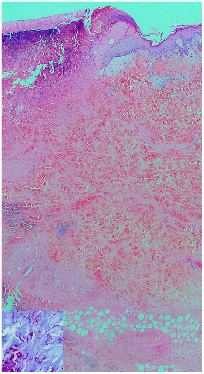
Figure 1. Skin biopsy from patient A after diagnosis of BU.

Skin biopsy (×2.5 magnification) showing ulceration and extensive undermining necrosis of the dermis and subcutaneous fat with minimal inflammation, typical of B.U. Insert, Wade Fite stain (×40 magnification) showing numerous acid fast bacilli.
