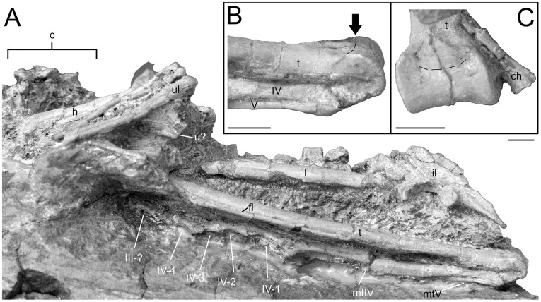
Figure 6. Hindlimb details of Mei long DNHM D2514.
A, articulated left hindlimb in lateral view; B, distal tibia and metatarsals IV and V of left hindlimb. Arrow and dashed line indicate caudodorsal edge of articular surface; C, distal end of tibia in caudodistal view. Dashed line as in B. Abbreviations as in Figure 1. Scale bars equal 0.5 cm.

