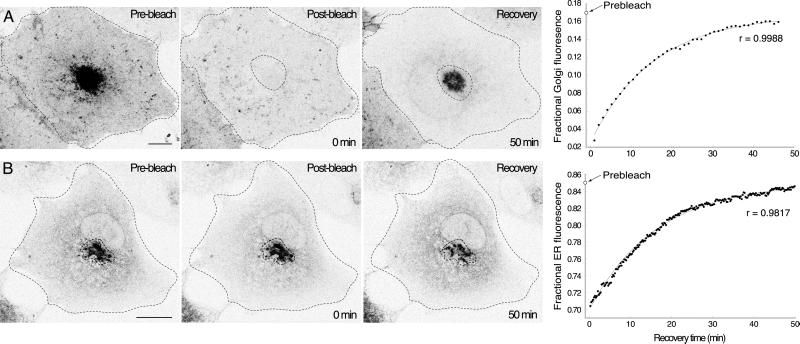
Figure 7.
Organelle-specific bleaching determines the kinetic parameters of VIP36-SP-GFP exchange between the ER and Golgi. See the online data for bleach parameters and controls. (A) Golgi-specific bleach experiments directly measure the transfer of fluorescence from the ER to the Golgi. The Golgi region defined by the dotted line was selectively bleached in a COS cell transiently expressing VIP36-SP-GFP. Panels show before, immediately after, and 50 min after the bleach for a representative experiment. Images are brightest point projections through a series of confocal sections over the entire depth of the cell. The recovery of fractional Golgi fluorescence for the cell shown is plotted on the far right (see MATERIALS AND METHODS), and the fit of the kinetic model is indicated by the light gray line through the data. The prebleach fractional Golgi fluorescence is indicated on the y-axis. After 50 min of recovery the cell appears dimmer because fluorescence was removed by the bleach (online data). Bar, 10 μm. (B) ER-specific bleach experiments directly measure the transfer of fluorescence from the Golgi to the ER. The ER region was selectively bleached in a COS cell transiently expressing VIP36-SP-GFP. Panels show before, immediately after, and 35 min after the bleach for a representative experiment. Images are brightest point projections through a series of confocal sections over the entire depth of the cell. The recovery of fractional ER fluorescence for the cell shown is plotted on the far right (see MATERIALS AND METHODS), and the fit of the kinetic model is indicated by the light gray line through the data. The prebleach fractional ER fluorescence is indicated on the y-axis. Bar, 10 μm.

