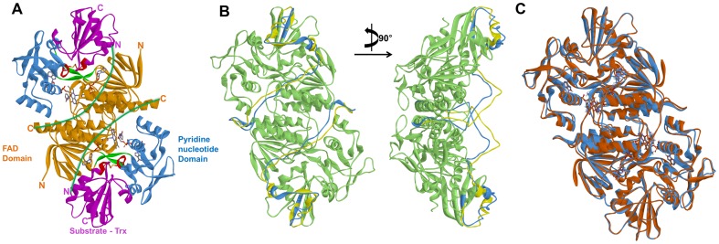
Figure 2. Three dimensional structures of template and the two proteins with refined linker region by 20 ns MD simulation.
(A) The template E. coli TrxR-Trx complex representing FAD (orange), PD (blue), and Trx (magenta) domains and functional regions (red) along with the missing linker region (green). (B) Showing the modeled missing linker region for the AtNTR_CC before (yellow) and after (light blue) 20 ns MD simulation. (C) Showing the superimposed homology modeled structures of AtNTR_CC (light blue) and AtNTR_AC (light brown), indicating the functional domains in different colors along with cofactors.

