
Figure 3. Confocal images of the stereocilia and prestin immunoactivity, and the pattern of Atoh1-EGFP expression in the organ of Corti.
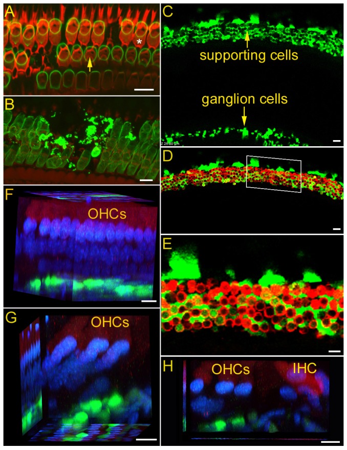
A: Stereocilia bundles in one cochlear location. The hair bundle was labeled with rhodamine-phalloidin (in red). The “V”-shaped OHC bundles were missing in the outmost (third) rows of OHCs. One bundleless OHC is indicated by an asterisk. One “V”-shaped hair bundle is marked by a yellow arrow. Prestin immunoactivity was labeled in green. Scale bar: 10 µm. B: Z-axis stack image of prestin immunoactivity at another cochlear location. Scale bar: 10 µm. All the images in A and B were obtained from cochleae 10 days after noise exposure. C: Confocal image of Atoh1-expression in a cochlea. D: Composite images of hair cells (labeled with myo7a antibody, in red) and EGFP-positive cells (from the same location as shown in panel C). The images were acquired seven days after Ad.Atoh1-EGFP inoculation in the noise-damaged cochlea (second turn). A magnified image of the area marked by white lines is presented in panel E. Scale bar: 20 µm for C and D. E: High magnification image of Atoh1 and myo7a expression in the organ of Corti. Scale bar: 10 µm. F: Confocal image obtained from optical sectioning from a basal turn location at 3 days after Atoh1 treatment. Hair cells were labeled with myo7a (in red) and the nuclei were labeled with DAPI. Most preparations examined at 3 days after transfection showed weak or no expression of EGFP in the organ of Corti. For those that expressed EGFP, the expression was in the area of Deiters' cells. G, H: Confocal image using optical sectioning from a basal turn location at 7 and 14 days after Atoh1 treatment. Scale bars (F,G, H): 10 µm.