
Figure 5. Stereocilia bundle counts in the normal (without noise exposure), Atoh1-treated and untreated cochleae.
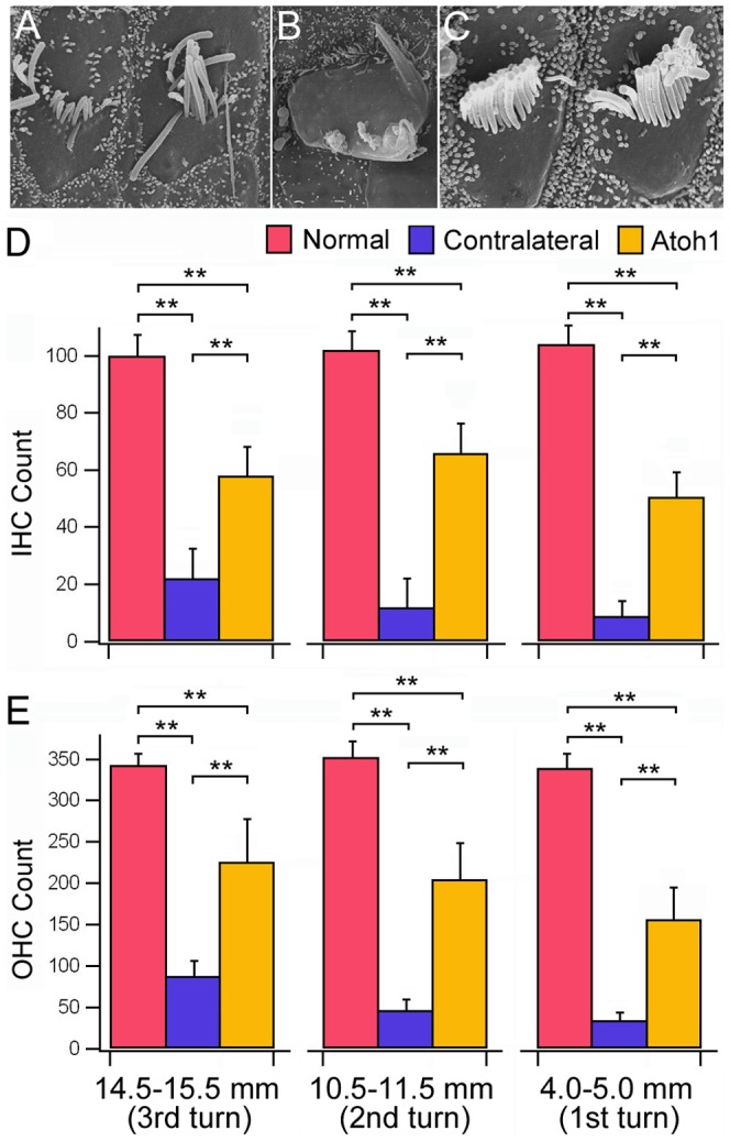
A–B: Examples of damaged bundles. C: Examples of normal-looking bundles. Scale bar represent 5 µm for all images in panels from A to C. D, E: Counts of IHC and OHC bundles in three different cochlear locations, each 1 mm in length. Three cochleae were used for each group (normal, Atoh1-treated, and untreated). Status of the bundles was determined by visual inspection of the bundles under scanning electron microscope. Hair bundles with no clear signs of trunction, fusion, or folding were included in the bundle count. Student's t-test was used for statistical analysis. Two asterisks represent statistical significance with a p-value less than 0.01.