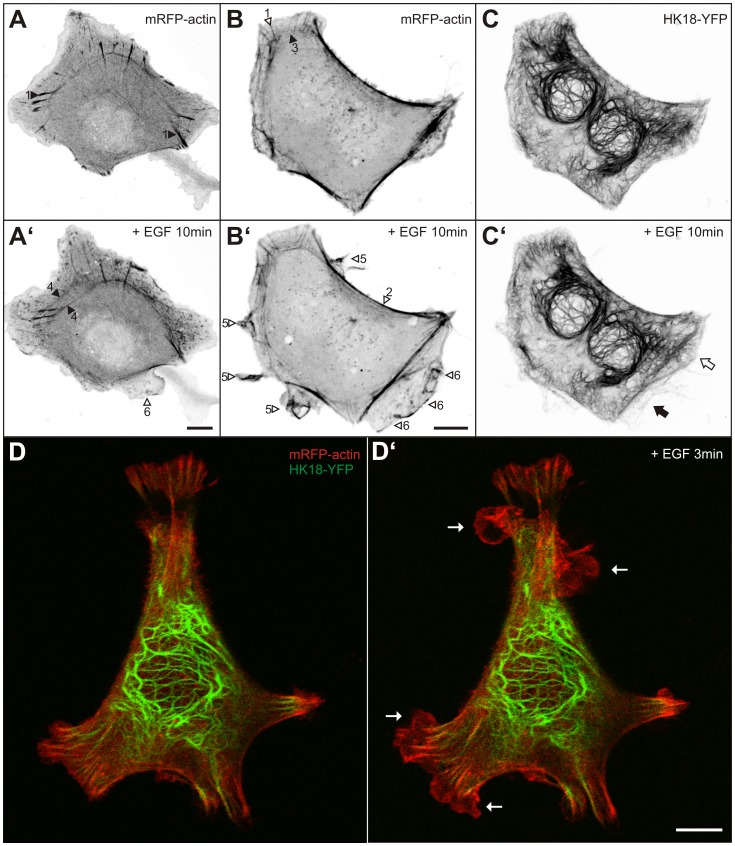
Figure 2. EGF induces actin-rich and keratin-free ruffles and lamellopodia.
(A–B’) Fluorescence microscopy of MKN1 cells producing mRFP-actin before (A, B) and 10 min after addition of 30 ng/ml EGF (A’, B’). Focal adhesion-associated stress fibers (1), submembranous stress fibers (2), transverse fibers (3) and transverse dorsal arcs giving rise to centrally located circular bundles (4) can be distinguished. In addition, cortical enrichment of actin is noticeable in ruffles and lamellipodia (5) and in lamellar extensions (6). Note that prominent actin stress fibers persist, actin-rich ruffles, lamellipodia, and lamella are generated, and that circular actin bundles translocate toward the cell center in the presence of EGF. (C–C’) The binucleated cell shown in (B, B’) also expressed HK18-YFP. Note that only the older lamella is positive for HK18-YFP fluorescence (white arrow in C’) whereas the newly-formed lamella below is not (black arrow in C’). (D–D’) The overlay images show a live MKN1 cell that is labeled with HK18-YFP (green) and mRFP-actin (red) before (D) and 3 min after addition of 5 ng/ml EGF (D’; complete sequence in Video S2). Note again the appearance of multiple keratin-free ruffles (arrows) upon EGF treatment. Bars, 10 µm.

