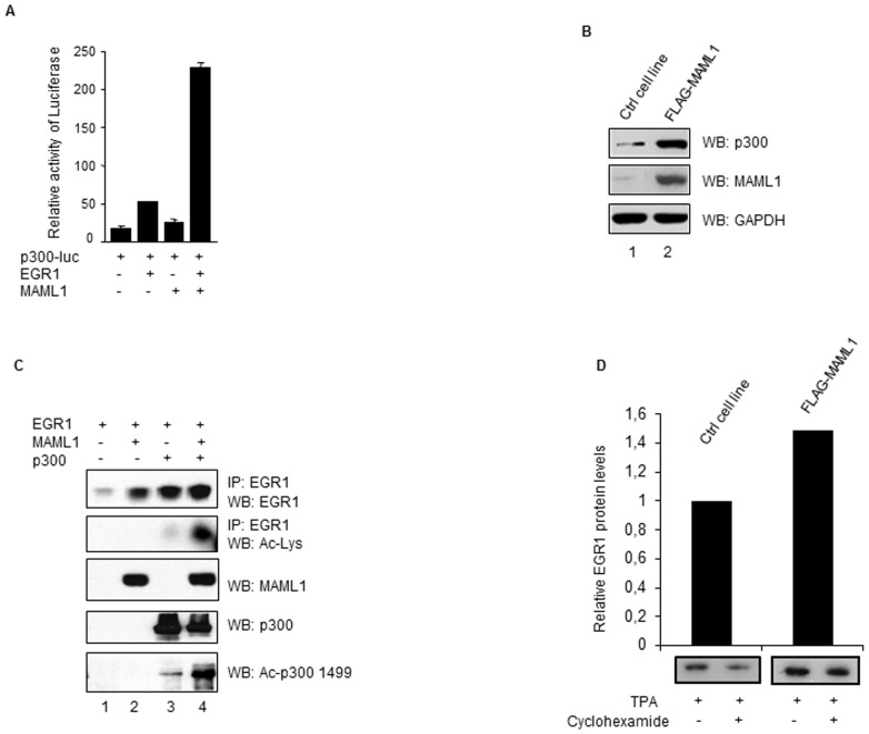
Figure 3. Activity of the p300 promoter is increased by MAML1.
(A) HEK-293 cells were co-transfected with a p300-luc reporter, EGR1 and MAML1, then cultured in the presence of TPA to induce expression of EGR1. The data is presented as mean ± SD. (B) Whole cell extracts were prepared from FLAG-MAML1 transfected and control HEK-293 cells (Ctrl) and MAML1, p300 and GAPDH were detected by Western blotting. (C) Whole-cell extracts were prepared from HEK-293 cells transfected with vectors expressing FLAG-EGR, MAML1 and p300; FLAG-EGR1 was immunoprecipitated using an antibody recognizing the FLAG epitope. The proteins were separated by SDS-PAGE and detected by Western blotting using antibodies recognizing EGR1, acetylated lysines in EGR1, MAML1, p300 and acetylated lysine1499 of p300. (D) HEK-293 cells stably expressing FLAG-MAML1 and HEK-293 control cells were cultured with TPA (2 h) in the presence or absence of cyclohexamide (1) and EGR1 protein expression was analyzed by Western blotting using an antibody recognizing EGR1. In the graph, EGR1 protein expression in the MAML1 stable cell line is expressed relative to control cells treated with cyclohexamide. The ratio of band intensities before the addition of cycloheximide and after 1 h of treatment with cycloheximide was determined with Image J.

