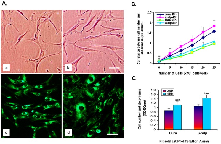
Figure 1. Fibroblast characterization: FSP-1 protein expression by immunofluorescence staining and cell proliferation assay in dura and scalp. A.
The morphology of postmortem fibroblast cells generated from (a) dura and (b) scalp. Cultured cells from both sources macroscopically looked similar to what is seen in living skin fibroblast cells, with more enriched cytoplasm and spindle-shaped nuclei under phase-contrast microscopy. Cells from (c) dura and (d) scalp express cytoplasmic Fibroblast Specific Protein-1 (FSP-1) (green). Original scale bars = 35 µm. B. Results from cell proliferation assay in 8 fibroblast cell lines (dura and scalp from 4 individuals) in five different densities. Cell viability was determined in 24 hrs and 48 hrs by WST-8 assay. Values are the mean of results from six wells. Bars ± SE. Scalp fibroblast cell lines grew 1.27-fold faster in the same period than dura fibroblast cells. C. Differences in cell proliferation between scalp and dura by one-way ANOVA; scalp cell growth was significantly more rapid than dura cell growth at 24 hr and 48 hr intervals [F (1, 46) = 12.94, p<0.008].

