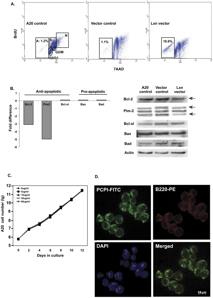
Figure 4. Ectopic Lxn expression increases apoptosis of A20 cells.
(A) Flow cytometric analysis of cell cycle and apoptosis in cultured A20 cells. A20 cells without (A20 control) or with empty (vector control) or Lxn-containing vector (Lxn vector) were cultured for 21 days. BrdU and 7AAD staining and flow cytometric analysis was used for detecting different phases of the cell cycle (G0/G1, S, G2/M) and apoptotic (A) and necrotic (N) population. A representative flow cytometric file from Day 5 of culture is shown. (B): Down-regulation of anti-apoptotic genes by Lxn overexpression. Apoptosis PCR arrays were performed on A20 cells infected with empty or Lxn-expressing vectors. Left panel shows the fold changes in the mRNA expression of several selected genes (as indicated in X axis) in Lxn-overexpressing cells compared to the control. Western blot confirms the decreased expression of Bcl-2 and two isoforms of Pim-2 at the protein level in Lxn-overexpressing cells (arrowhead). The other apoptosis-related genes, such as Bcl-xl, Bax and Bad, did not show significant difference in mRNA and protein expression. (C) The growth curve of A20 cells treated with graded doses of potato carboxypeptidase inhibitor (PCPI). No significant difference is detected between the control and any of the doses of PCPI. (D) Internalization of PCPI to cytosol of A20 cells. A20 cells were cultured with fluorescein isothiocyanate (FITC) labeled PCPI. Shown is a three-color micrograph with PCPI in green (FITC), the B220 lymphoid cell surface marker in red (phycoerythrin, PE), the nucleus in blue (DAPI), and merged image of red and green as indicated.

