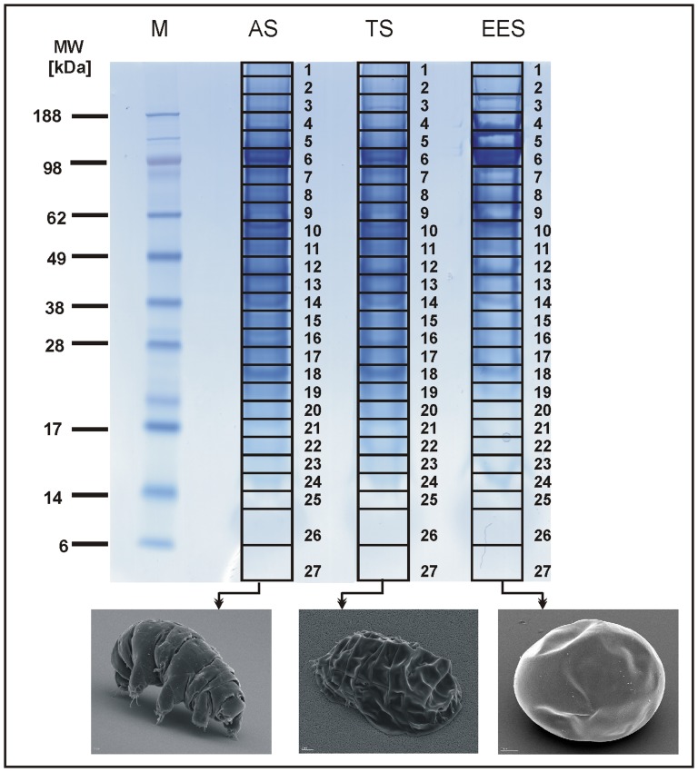
Figure 1. Separation of protein lysates of tardigrades in three different states by one-dimensional polyacrylamide gel electrophoresis.
Lane 1: Rainbow molecular weight marker. Lane 2: Protein extract of adult tardigrades in active state (AS). Lane 3: Protein extract of adult tardigrades in tun state (TS). Lane 4: Protein extract of tardigrades in early embryonic state (EES). Bottom. SEM-images of M. tardigradum in the corresponding states.

