Figure 1.
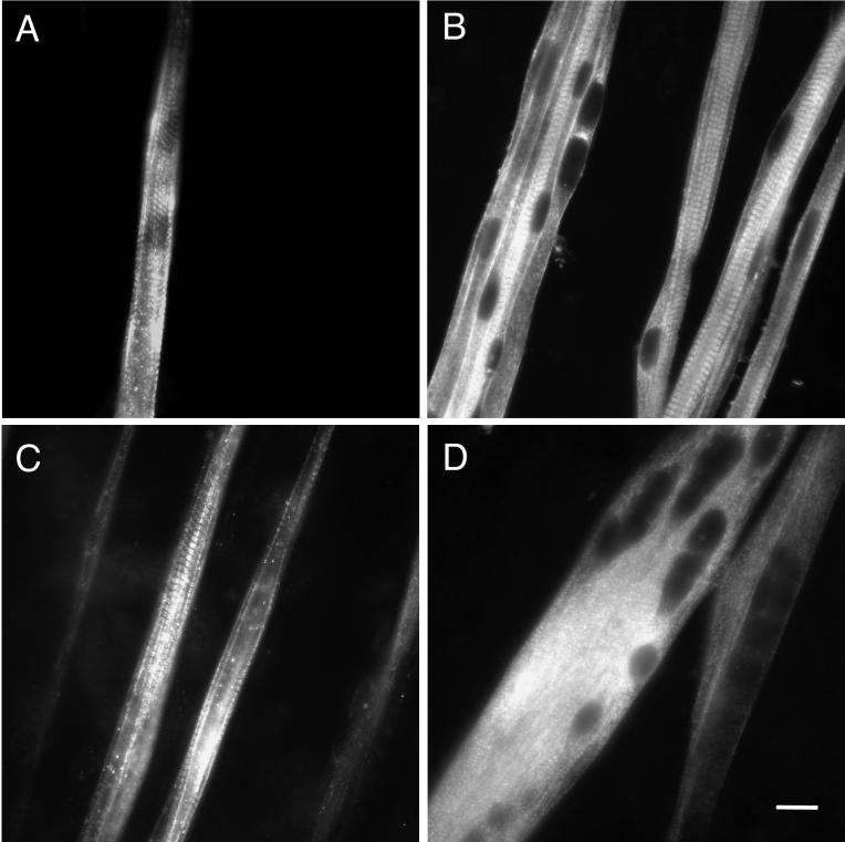
Protein expression of MHC isoforms in primary cultures of rat myotubes. Myoblasts were isolated from embryonic day 21 rats and cultured for 12–13 d, during which time they fused into multinucleated myotubes. They were fixed and immunostained as described in MATERIALS AND METHODS. (A) Typical view of myotubes stained for slow MHC, showing nonuniform staining and the presence of cross-striations. (B) Myotubes stained for embryonic MHC. Myotubes exhibited well organized cross-striations and often had peripheral nuclei. (C) Myotubes stained for fast/neonatal MHC illustrating the onset of sarcomeric organization and variations in staining intensity. (D) Culture treated with 1.5 μM TTX and stained for embryonic MHC. Myotubes were broader, had a paucity of cross-striations, and more central nuclei. Bar, 20 μm.

