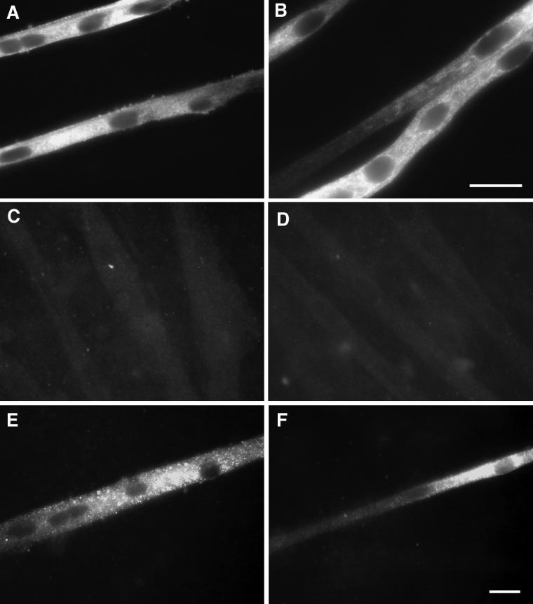
Figure 3.
Protein expression of slow MHC in myotube cultures. Cells were cultured as described in Figure 1 and immunostained with an antibody against slow MHC. (A and B) Culture transfected with CN*, showing widespread, intense punctate staining, central nuclei, and a scarcity of cross-striations. (C) Culture administered CSA. No staining above background was detected. (D) Culture transfected with CN* and administered CSA. No staining above background was detected. (E) Culture transfected with CN* and administered tetrodotoxin. Staining was decreased compared with transfection with CN* only. (F) Culture transfected with CN* and VIVIT. Staining was absent, or the intensity was greatly decreased compared with transfection with CN* only, except in limited regions of a few myotubes. Bars, 20 μm (bar in B refers only to that panel).

