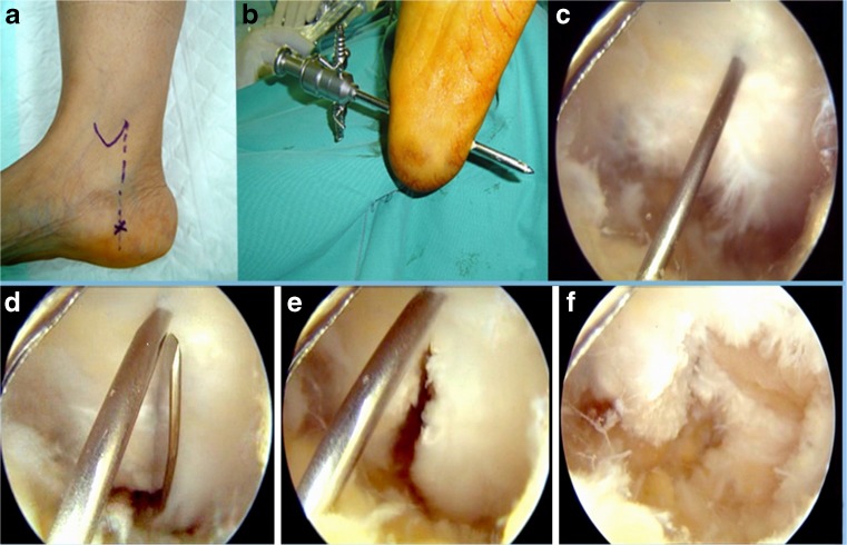
Fig. 2.
(a) Intra-operative photograph showing the landmarks of the medial portal. (b) Endoscopic view showing the shiny fibers of the plantar fascia and a needle acting as a landmark for the middle of the plantar fascia. (c) Endoscopic view showing a standard scalpel blade No. 11 introduced through the medial portal. (d) Endoscopic view showing the full thickness of the medial half of the plantar fascia divided into two leaflets. (e) Endoscopic view after debridement of the posterior leaflet. (f) Intra-operative photograph showing the final appearance of the medial portal at the end of the operation

