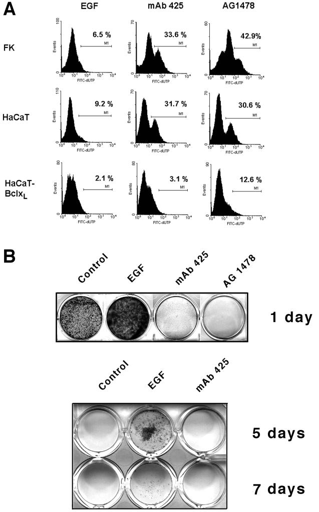
Figure 1.
EGFR-dependent survival and viability of keratinocytes in forced suspension culture. (A) TUNEL staining of normal neonatal keratinocytes (FK), HaCaT keratinocytes, and HaCaT cells overexpressing Bcl-xL–maintained in suspension for 24 h in the presence of EGF (10 ng/ml), mAb 425 (10 μg/ml), or AG1478 (10 μM) as indicated; values represent the cell fractions stained with FITC-dUTP relative to controls, i.e., attached HaCaT cells. (B) Clonal growth of HaCaT cells recovered after 1, 5, and 7 d of suspension culture in different medium conditions as indicated, further propagated on tissue culture-treated plastic for 5 d, and stained with crystal violet.

