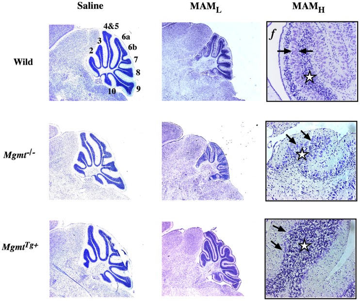
Figure 1.
Cytoarchitecture of the cerebellum of wild-type and DNA-repair mutant mice treated with MAM acetate. Light micrographs of representative areas from cresyl violet-stained parasagittal sections (10 μm) of the postnatal 22-day-old cerebellum from C57BL/6J (wild), Mgmt−/−, or MgmtTg+ treated on postnatal day 3 with a single injection of saline (left panels) or MAM 325 μmol, s.c. (center panels). Higher magnification of the cerebellum from wild-type or DNA repair mutant mice treated with MAM (right panels). MAML, low magnification (3.85×), MAMH, high magnification (77×), f, folia. Arrows indicate disorganization of Purkinje cell layer and stars denote reduced density of neurons in granule cell layer. Mgmt: gene coding for O6-mG-methyltransferase (MGMT), which is knocked out in Mgmt−/− and overexpressed in MgmtTg+ mice (modified from Kisby et al., 2009).

