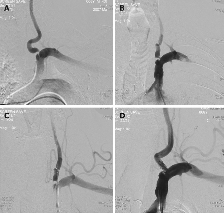
Figure 2.

Classical approach for vertebral artery stenting. A-C: Left subclavian angiography posterior-anterior (PA) view shows ulcerated short segment significant stenosis in the origin of the left vertebral artery (A), placement of the microguidewire to the left vertebral artery and microguidewire to the subclavian artery as a guidewire (B) and correct placement of the coronary balloon-expandable stent in the stenosed segment (C); D: Post-stent angiography shows good opposition of the stent and well opening of the stenosed segment.
