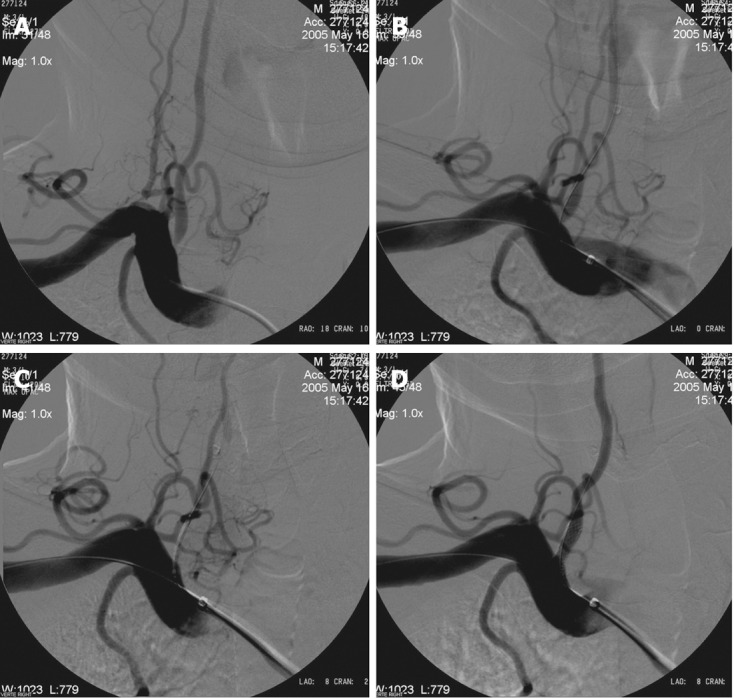
Figure 3.

Buddy wire technique for tortuous subclavian arteries. A: Right subclavian angiography posterior-anterior (PA) view shows significant concentric stenosis of the right VA origin; B: Right subclavian angiography shows placement of the stent to the right vertebral artery (VA) and one microguidewire to the subclavian artery as a buddy wire; C: Right subclavian angiography after placement of the stent shows temporary occlusion of the right VA and correct placement of the stent; D: Right subclavian angiography after opening of the stent shows good anatomical result.
