
Figure 4.
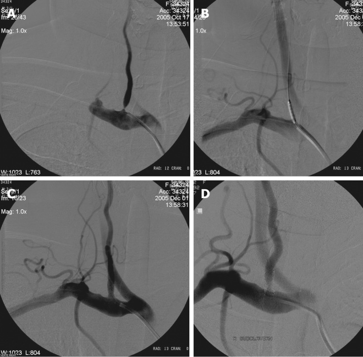
Radiological follow-up. A: Right subclavian angiography posterior-anterior (PA) view shows significant stenosis of the right vertebral artery origin; B: Placement of the coronary balloon-expandable stent in the stenosed segment; C: After opening of the stent angiography shows well opposition of the stent and lack of residual stenosis; D: Eighteen months control angiography shows patency of the right VA origin and stent.