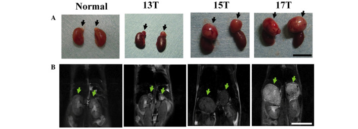
Figure 1.
Images of adrenal tumors in a transgenic mouse by MRI. (A) Gross appearances and (B) T2-weighted coronal images from a transgenic mouse at the ages of 13, 15, and 17 weeks (13T, 15T, and 17T, respectively) and normal adrenal glands from a non-transgenic littermate at the age of 13 weeks. Arrows indicate the adrenal glands in the non-transgenic littermate and adrenal tumors in a transgenic mouse. Bars, 10 mm.

