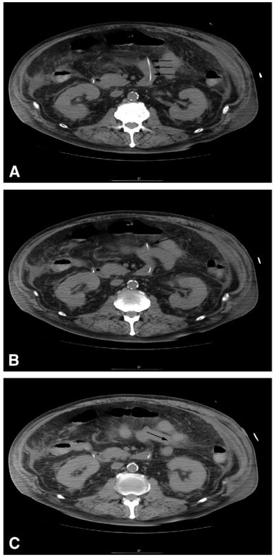
Figure 2.
Subsequent CT scan with jejunal tube extension in progressively distal small bowel. A, Jejunal tube (arrows) traveling anteriorly in the small bowel. B. Distal tip of the jejunal tube (arrow) terminating at a bend of the small bowel, producing mucosal tenting. C, The bowel traverses toward the patient’s left (arrow) without the jejunal tube.

