
Figure 3.
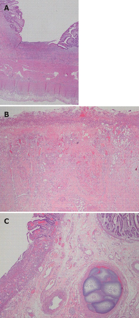
Surgically resected jejunum. A: Full thickness small bowel section showing ulcerated small intestinal mucosa; B: Ulcer bed showing acute and chronic inflammation, granulation tissue and overlying fibrinopurulent exudate, no granulomas are seen; C: Intravascular embolization material in submucosal blood vessel.