Figure 1.
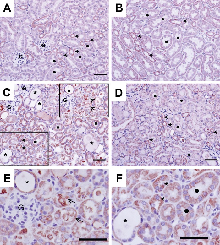
COX 1 immunohisto-chemistry in mouse kidney 72 hr after cisplatin treatment. Panels A and B: control sham-treated mouse kidney. Extensive immunostaining (red stain) is observed in both proximal and distal tubular epithelium; staining is mainly in the basolateral surface of the epithelia. In the cortex (Panel A), distal tubules (arrowheads) are more extensively stained for COX 1 than are proximal epithelium (denoted by ●). In the medulla (Panel B), distal tubules (arrowheads) also exhibit more intense staining than S3 segments of proximal tubules. Panels C, D, E, and F: Cisplatin induced profound histopathological renal injury, desquamation of epithelial cells evidenced by protein cast in the tubular lumen (arrows), and dilation of the tubules (denoted by *). COX 1 expression was mildly decreased in damaged tubular epithelial cells (Panel E), but immunostaining is maintained at a relatively normal distribution in spared epithelium (Panel F). Bars: A, B, C, D: 50 µm.

