Figure 6.
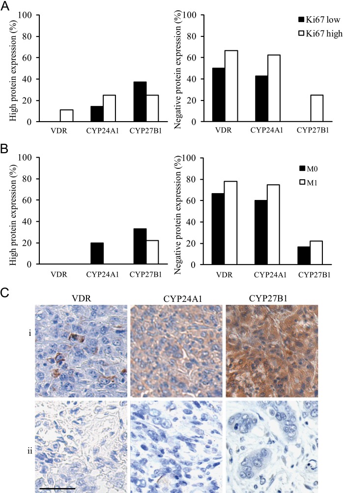
“High” (left histogram) and “negative” (right histogram) protein expression profile of vitamin D receptor (VDR), CYP24A1, and CYP27B1 in anaplastic thyroid cancer (ATC) subdivided according to their Ki67 status (low, high) (A) and according to the presence of metastasis (M0, M1) at diagnosis (B) and representative photographs of a triple-negative (absent VDR, CYP24A1, and CYP27B1) and triple-positive staining in ATC subdivided according to their Ki67 status (C). (A) More cases of absent VDR, CYP24A1, or CYP27B1 staining were present in ATC cases with a Ki67 “high” status compared with a Ki67 “low” status. (B) Also, more cases of absent VDR, CYP24A1, or CYP27B1 staining were found in ATC M1 compared with ATC M0. (C) Representative photographs of two ATC cases with either a triple-positive or a triple-negative staining for VDR, CYP24A1, and CYP27B1 in, respectively the (i) Ki67 low and (ii) Ki67 high ATC subgroup. Scale bar: 50 µm.

