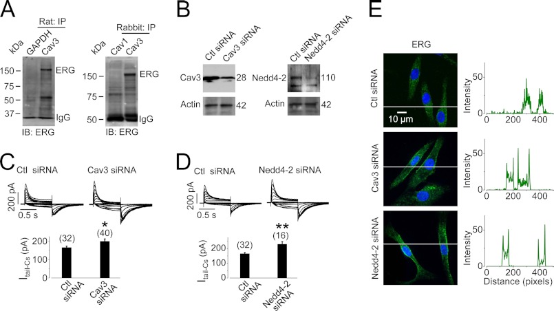
FIGURE 8.
Cav3 interacts with IKr in ventricular tissues. A, Cav3 interacts with native ERG channels. ERG was detected in proteins precipitated with an anti-Cav3 antibody from membrane proteins extracted from both rat (n = 4) and rabbit ventricular tissues (n = 5). GAPDH or Cav1 was used as negative control. B, knockdown of Cav3 or Nedd4-2 expression is shown in cultured neonatal rat ventricular myocytes. C and D, knockdown of Cav3 (C) or Nedd4-2 (D) increases IKr in cultured neonatal rat ventricular myocytes. Families of Cs+-mediated IKr were recorded 24–48 h after siRNA transfection. The numbers in parentheses above the bars indicate the number of cells tested. *, p < 0.05; **, p < 0.01 versus control (Ctl) siRNA. E, knockdown of Cav3 or Nedd4-2 retains cell surface ERG proteins in cultured neonatal rat ventricular myocytes after 4-h culture. The intensities of green signals along the lines across the cells are plotted against pixel distances. Whereas the signal is evenly distributed in control siRNA-transfected cells, it peaks at the plasma membrane in Cav3- or Nedd4-2 siRNA-transfected cells.

