
FIGURE 5.
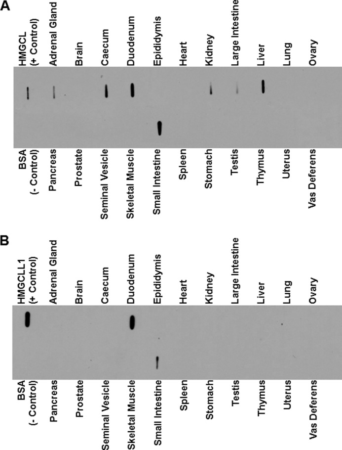
Endogenous tissue expression of HMGCLL1. Protein (10 μg) of clarified organ lysates from the indicated rat tissues was applied to nitrocellulose using a slot-blot manifold. Negative control lanes contained BSA (1 μg); positive control lanes contained 1 ng of purified HMGCL or HMGCLL1. Blocked, peroxidase suppressed blots were incubated separately with anti-HMG-CoA lyase (A) or anti-HMGCLL1 antibodies (B). The blots were incubated with goat anti-rabbit IgG horseradish peroxidase-conjugated secondary antibody and developed using enhanced chemiluminescence. As demonstrated in Fig. 2, the antibody against HMG-CoA lyase recognizes both mitochondrial HMG-CoA lyase and HMGCLL1, whereas the antibody against HMGCLL1 does not recognize mitochondrial HMG-CoA lyase.