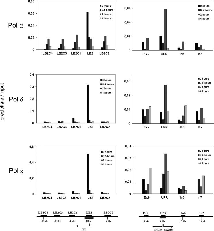
FIGURE 2.
Time course of the association of origin of replication DNA within the LB2 gene (left panels) and the UPR of the MCM4 gene (right panels) with nucleoprotein complexes isolated by immunoprecipition of S phase Pol α, δ, and ϵ. The regions analyzed by qPCR are indicated in the panels and their locations are shown below the panels, the LB2 origin in the left panel and UPR origin in the right panel. The origins are shown as gray boxes. Black bars represent regions that were amplified by quantitative PCR. The genomic organization and the location of the PCR products are identical to Fig. 1. The cells were synchronized by double thymidine block to early S phase (0 h) and released to proceed in S phase (0.5, 2, and 4 h). Nucleoproteins derived from CsCl centrifugation were sonicated and digested with micrococcal nuclease as described under “Experimental Procedures.” Soluble nucleoproproteins, 1 mg/500 μl, were taken for immunoprecipitation with the cognate antibodies, the protein was digested with proteinase K, and DNA isolated. 5 to 10 ng of DNA was obtained from the immunoprecipitate, 1/20 was used for quantitative PCR (precipitate). DNA from soluble nucleoproteins representing genomic DNA was isolated and treated in the similar manner, except that the immunoprecipitation step was omitted, and analyzed by quantitative PCR (input). The values are mean values from two distinct experiments.

