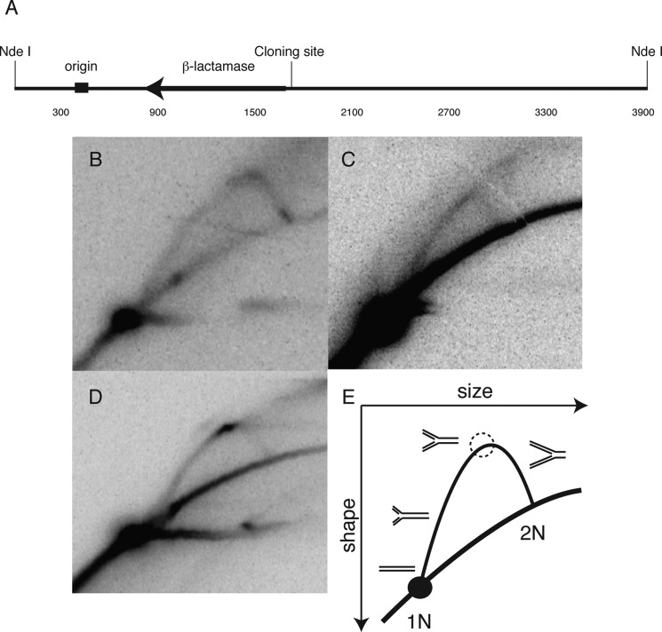
FIGURE 2.
In vitro replication demonstrates a polar replication blockade. A, map of the SV40 origin of replication in relation to the cloning site of the PKD1 mirror repeat sequences and the NdeI restriction endonuclease cut sites in the pZ189-based vector. B, neutral-neutral two-dimensional replication gel revealing a smooth Y arc of replication intermediates for the vector alone. C, a smooth Y-arc similar to the replication pattern of the vector alone is seen when leading strand synthesis encounters the Pu-rich template. D, a strong pause is seen when the Pu-rich sequence is in the lagging strand template. The horizontal signal extending from the 1N spot is commonly seen in plasmid replication (93). E, diagram explaining the results in panel D, depicting the increase in signal due to fork stalling (dashed circle, on arc of Y-intermediates).

