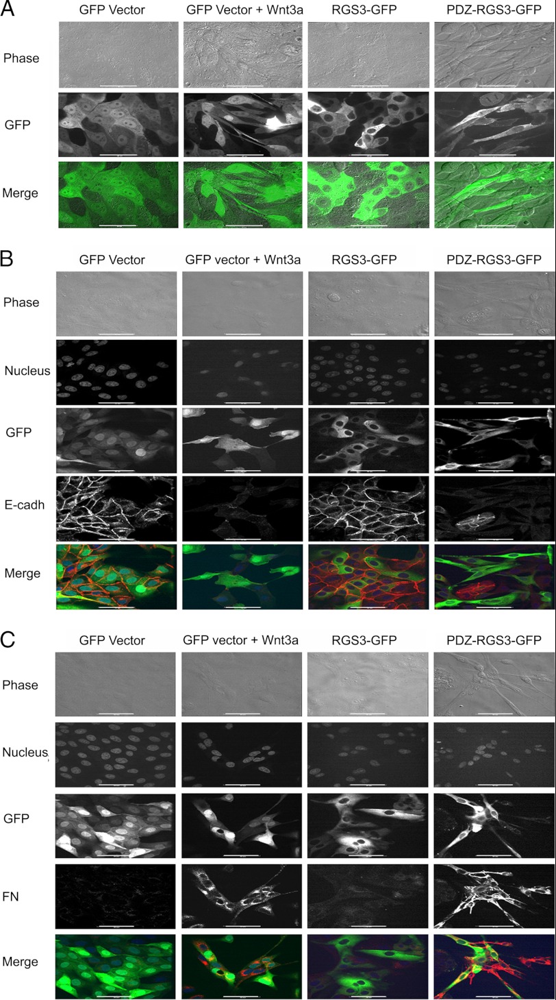
FIGURE 7.
PDZ-RGS3 expression mimicked Wnt3a treatment of MDCK cells and triggers EMT. A, Wnt3a treatment or PDZ-RGS3 expression, but not RGS3 expression, caused MDCK cells to lose their epithelial characteristics. Stable MDCK transfectants of GFP vector, RGS3-GFP, or PDZ-RGS3-GFP were generated. Images of live cells are shown. The indicated cells were exposed to Wnt3a (20 ng/ml) overnight. B, Wnt3a and PDZ-RGS3 expression decreased E-cadherin expression. The indicated cells were immunostained for E-cadherin (red). Phase contrast, nucleus (far red), green fluorescent, and red fluorescent images were acquired. An E-cadherin (red) with GFP (green) merge is also shown. C, Wnt3a treatment or PDZ-RGS3 expression increased fibronectin (FN) expression. The cells stably transfected with indicated constructs were fixed and immunostained for fibronectin. Phase contrast, nucleus (far red), GFP (green), and fibronectin (red) images were acquired. A merged image between GFP and fibronectin is shown in the bottom panels. Scale bar, 20 μm.

