FIGURE 3.
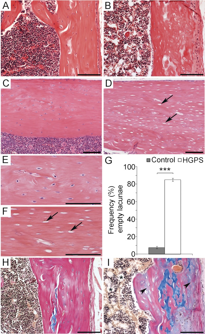
Progerin expression during osteoblast development results in loss of osteocytes and mineralization defects. The histological analysis of the femur of wild-type (A, C, E, and H) and HGPS mice (B, D, F, and I). Hematoxylin & Eosin staining on femur sections (A–F). In sections from wild-type mice viable osteocyte cells were detectable (C, and enlarged in E) while sections from HGPS mice showed a high frequency of empty osteocyte lacunae (arrow) (D, and enlarged in F). Quantitative histomorphometric analysis showed a significant increase of empty osteocyte lacunae in HGPS mice 85.1 ± S.E. 1.8% compared with wild-type mice 7.4 ± S.E. 1.4% (G). Unmineralized matrix was detected by Alcian blue & Van Geison staining (H, I) with cartilage stained in blue, mineralized tissue stained pink, and unmineralized tissue stained gray (arrowhead). Increased adipocyte infiltration of the bone marrow space was detected in HGPS mice only (asterisk) (I). Scale bars: (A, B, H, I) 50 μm (C–F) 100 μm. Values represent the mean ± S.E. (***, p < 0.001).

