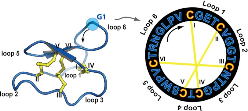
FIGURE 1.
Cyclotide structural information. Three-dimensional structure (Protein Data Bank code 1nb1) and sequence of the prototypic cyclotide kB1 are shown. Cyclotides are characterized by a cystine knot motif formed by three disulfide bonds, shown in yellow. The six Cys residues are labeled I–VI, and the residues between adjacent Cys residues are designated as loops 1–6. The direction of the peptide chain N-C is shown with a black arrow. G1 is highlighted as the starting point of the numbering scheme.

