FIGURE 6.
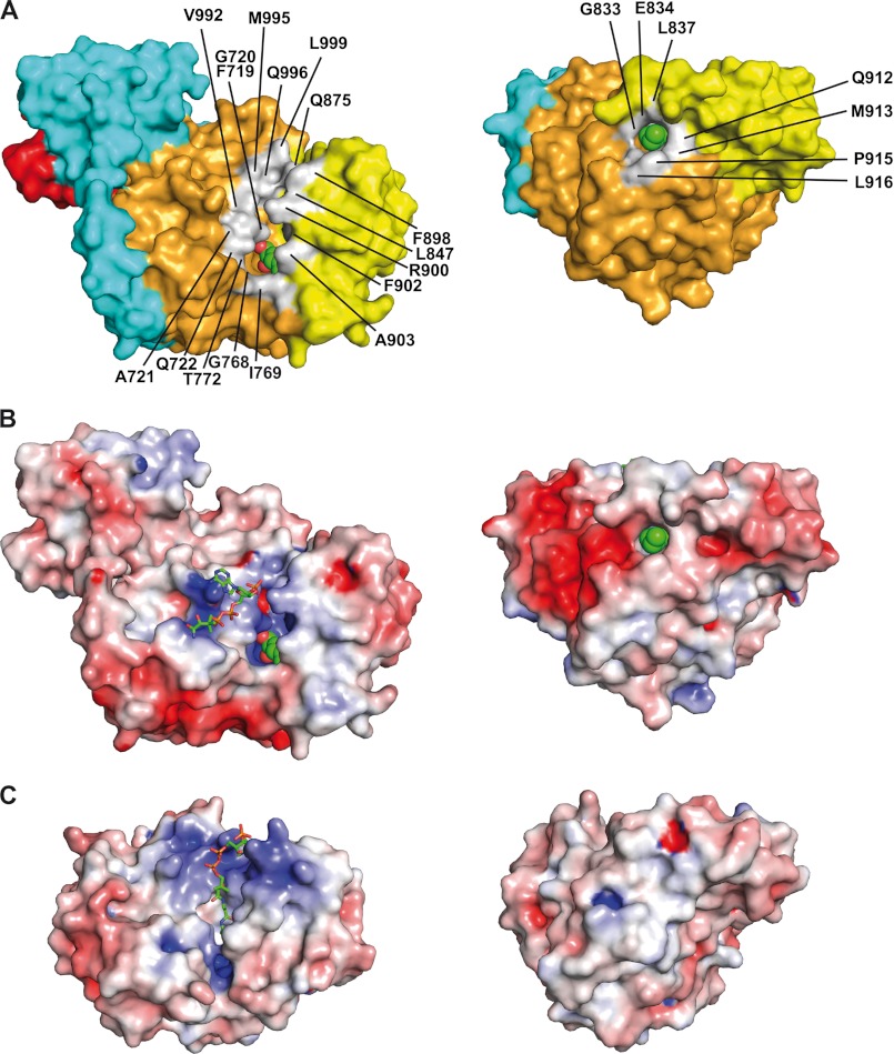
Topography of the active site and hydrophobic channel of AT52. A, molecular surface. B, electrostatic potential surface. Two perpendicular views are shown. Residues delineating the groove, at the apex of the active site, and the exit of the channel are colored in light gray and labeled. The catalytic serine and the carboxypalmitoyl chain are shown as spheres. C, electrostatic potential surface of E. coli MCAT (Protein Data Bank code 2G2Z). The CoA-SH moieties are shown as balls and sticks with green carbon atoms (nitrogen, blue; oxygen, red; phosphorus, orange; sulfur, yellow). Electrostatic potential is color-coded from red (−6 kBT/e) to blue (+6 kBT/e); white is neutral.

