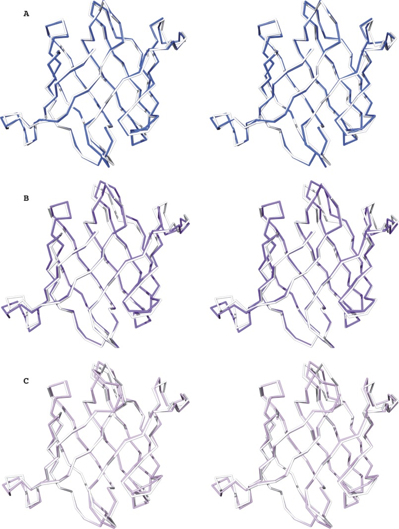
FIGURE 2.
Conservation of the OAAH fold. A–C, stereo views of best-fit Cα superpositions of OAA and PFA (A), OAA and the first domain of MBHA (B), and OAA and the second domain of MBHA (C), respectively. OAA, PFA, and the first and second domains of MBHA are colored in white, blue, purple, and light magenta, respectively.

