FIGURE 2.
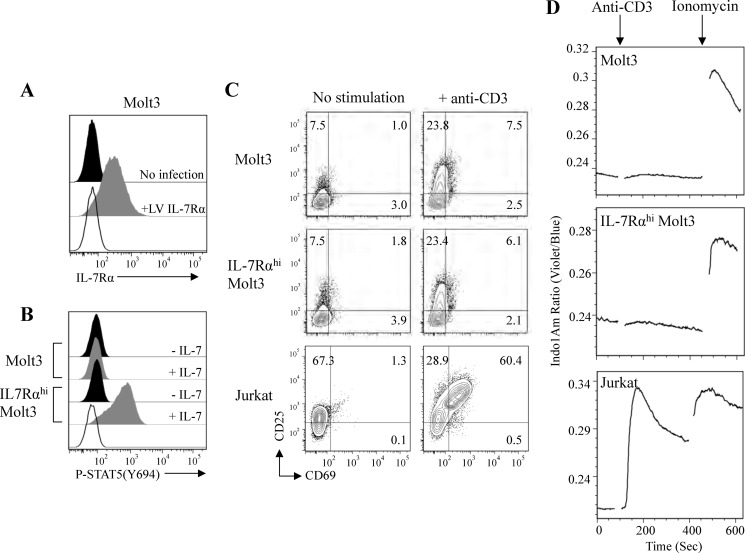
Analysis of IL-7 and anti-CD3 signaling in lentiviral vector-modified IL-7Rαhi Molt3. A, surface expression of IL-7Rα on Molt3 (no infection) and lentiviral vector-modified IL-7Rαhi Molt3 (+LV IL-7Rα). B, STAT5 activation in response to 100 ng/ml IL-7 by Phosflow antibody staining and flow cytometry analysis. C, surface expression of activation markers CD25 and CD69 in Molt3, IL-7Rαhi Molt3, and Jurkat cells after overnight incubation with anti-CD3/CD28 beads. D, intracellular free calcium measurement in Indo-1/AM-loaded Molt3, IL-7Rαhi Molt3, and Jurkat cells in response to anti-CD3 antibody (clone HIT3a) and ionomycin.

