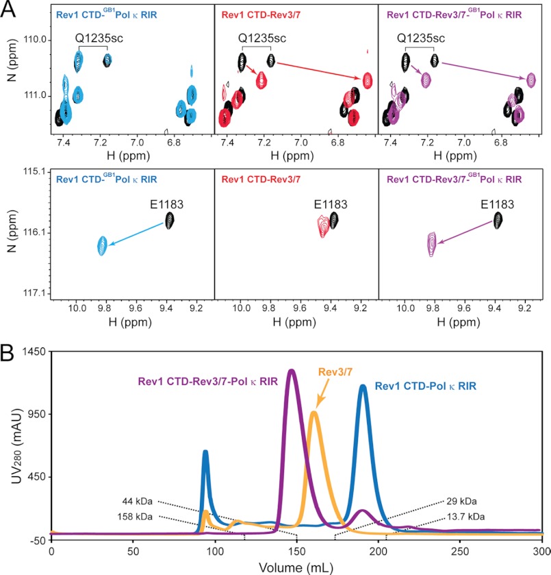
FIGURE 1.
Formation of a quaternary complex consisting of the Rev1 CTD, Rev3/7, and Pol κ RIR. A, 1H-15N HSQC spectra of the free Rev1 CTD (black) and the Rev1 CTD in the presence of an equal molar ratio of either the GB1-Pol κ RIR fusion protein (blue), Rev3/7 (red), or both (purple). B, FPLC traces of the Rev1 CTD-Rev3/7-Pol κ RIR complex (purple), the Rev3/7 complex (light orange), and the Rev1 CTD-Pol κ RIR complex (blue) separated using a HiPrep 26/60 Sephacryl S-200 HR column (GE Healthcare). The elution volumes of known protein standards are labeled.

